
 Sherolyn-Basement Bettas
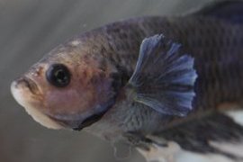
The white discolorations around the face and the swelling. See the grey areas along the back around the scale edges.
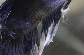
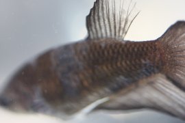
The entire area where the anal fin joins the body has the mycos. See the huge chunk of fin that has fallen off and the blacker areas of the body.
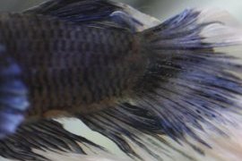
Again the blacker areas of the body have the mycos infection, Between the anal fin and body, and where the tail fin joins the body as well.
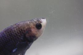
 You can see the grey areas or lesions on the top of the head, as well as under the eye and along the gill in the chin area. See the swelling and the white of the opposite side of the face.
Not the best photo but you can see all the patches of infection on the body of of this fish.
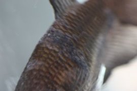
These are pretty advanced lesions on this fish. All the grey areas will eventually get darker like the larger area just before the dorsal fin.
Can see the very established areas of the mycos in front of the dorsal fin and further up on the head. And you can see all the white specs on the scales that are developing. The gill you see a lot of the white-grey areas that are also developing mycos.
Share this Post
latest post
-
 Betta fish Housing October 18, 2022
 -
 Betta fish spawning October 16, 2022
 -
 Interesting Facts about Betta fish October 14, 2022
 -
 AquaBid Betta fish October 12, 2022
 -
 Where do Betta fish originate from? October 10, 2022
 -
 Small Betta fish October 8, 2022
 -
 Betta fish without food October 6, 2022
 -
 Where are Betta fish found? October 4, 2022
 -
 Show Betta fish October 2, 2022